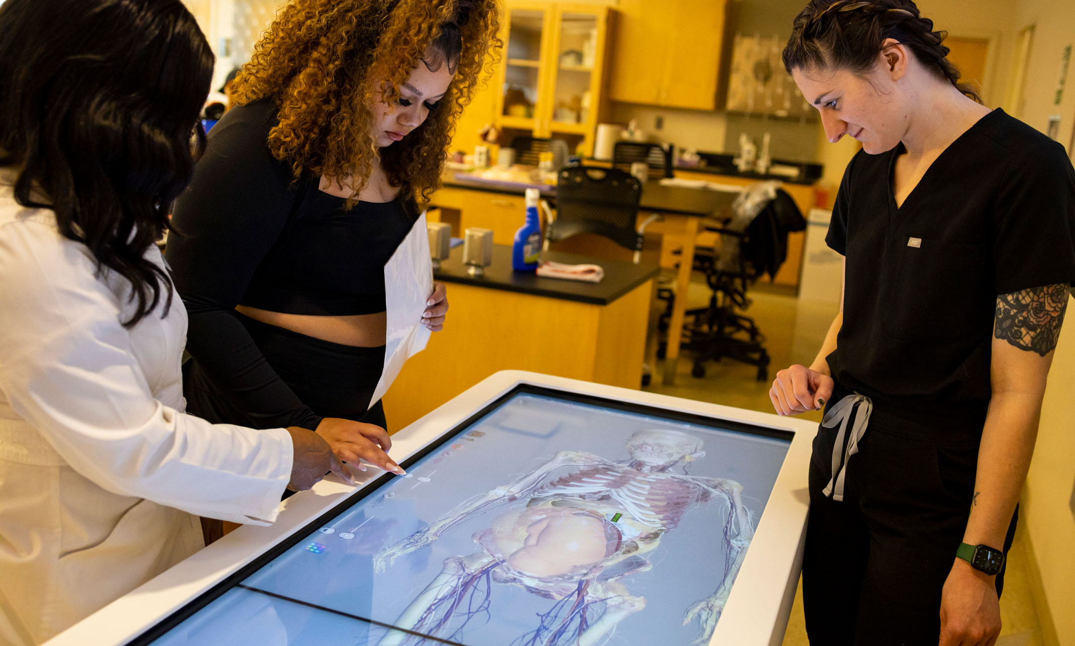
The Anatomage Table, a 3D anatomy visualization and virtual dissection tool, is about the size and height of a traditional hospital bed.
The top of the table features two integrated touch-screen glass monitors connected to a powerful computer housed in the base of the machine. At first glance, the screens look like any other computer interface, except the screen spans 7 feet.
The school acquired the table in the fall of 2019 through a state technology grant. Trainings were delayed because of the COVID-19 pandemic, and the school was able to start using the table in the Fall of 2021.
Shelley Hunter, Ph.D., a clinical assistant professor who teaches anatomy and physiology, is the resident expert on the inner workings of the impressive piece of technology. She said that even though Anatomage provided extensive training for faculty and staff, the technology was still daunting at first.
“At first I was really intimidated because the table is enormous, and it can do so many things,” Hunter said. “The students, though, are immediately comfortable with it. They just walk right up, and it doesn’t bother them since they were born into this age of technology.”
According to Hunter, the table presents a more “cleaned-up version” of the human body than students would experience in a traditional cadaver lab. With the Anatomage Table, if a student wants to see behind the kidneys, they just digitally move the organs out of the way. With a cadaver, those same kidneys would need to be removed by hand with considerable effort. For Hunter, the capabilities of the virtual anatomy table far outweigh the lack of tactile sensation that traditional dissection involves.
“For the cardiovascular unit we were studying, we pulled up the heart and it pulled up an EKG,” Hunter said. “Right beside it, and we could see the muscles, we could see the nervous system and the valves working while the ECG was going on and the students can relate the electrical with the mechanical.”
School of Nursing and Health Studies Dean Joy Roberts believes that UMKC is the only university in the Kansas City area offering this technology to its students. Roberts also knows first-hand that other area schools are interested in the table.
“I was on the board for North Kansas City School District. They called and said, ‘We hear you have this new machine and it’s really cool,’” Roberts said. “Leadership came to UMKC to check it out. They were so impressed they got one for the district, and plan to have them at all four of the North Kansas City high schools.”
Although the table itself is expensive, it’s still much cheaper than a cadaver lab.
“To operate a cadaver lab, you have to get people to donate their bodies,” Roberts said. “Then you have to store the cadavers, preserve them in formaldehyde and keep them secure. This table gives us access to cadavers without the terrible smell of formaldehyde burning our eyes.”
The school received the funding for the $80,000 table from a grant through the Nursing Education Incentive Program with the Missouri State Board of Nursing. According to Roberts, having access to cadavers, even if virtually, makes a world of difference for the students, providing them with more than a the traditional plastic model or picture in a book. For Roberts, those tried-and-true learning tools will always be useful, but the Anatomage Table provides a more complete picture.
Take, for example, the voice box. Hunter can zero in on that organ and remove related systems, such as circulatory or nervous systems, providing an unencumbered view of what students are focusing on for that particular lesson. The machine shines when Hunter takes a clear view of the voice box and rotates the entire organ.
The ability to do this is critical, because the muscles associated with the voice box are hidden behind the organ itself. Hunter can then show students the muscles that support the throat, the bone and cartilage, the Adam’s apple and the epiglottis, the flap that covers the windpipe. That full understanding is important for future nurses and their patients.
“Because most nurses will have to intubate a patient, we look at those muscles that control the opening and closing of that flap,” Hunter said. “We talk about what happens if any of these things are compromised. If the nerves or muscles aren’t functioning properly, the patient can die because there’s no way for air to get through.”
Hunter’s anatomy and physiology classes are comprised of pre-nursing students and health studies students. Classes consist of 20-35 students each, and Hunter has groups of five working at the table at one time. On any given review day, students can be found studying a unit, such as the cardiovascular system, watching the entire system work in real-time. Other groups quiz each other on terminology as they wait their turn at the table, where they’ll see a fuller picture of how circulation works.
Ashley Hanners is pursuing her bachelor’s degree in health studies, and she spent the past two semesters in Hunter’s class working with the Anatomage Table. Hanners came into the class from high school with some experience in dissection, which she said was helpful in working with table. There was one crucial difference between the virtual anatomy table and the mouse she dissected, however: the odor.
“The smell of chemicals and decomposition were potent,” Hanners said. “It made a lot of my high school classmates nauseous.”
Instead, Hanners appreciates that the table removed that distraction and enabled her to focus on diving deep into anatomy. She said that each person within her small group got better at identifying organs and systems, as well as dissecting and reconstructing those systems.
During a class in which Hunter reviewed the reproductive system, she showed students how uncomfortable pregnancy can be for expectant mothers. The students saw first-hand the pressure the baby puts on the surrounding organs, an exercise that elicited a variety of reactions from the students, including, “This is so cool,” “That is kind of scary,” and “You can see the baby’s heart beating!”

